Protocols

The standard procedures and protocols for the IU School of Medicine Center for Electron Microscopy (iCEM) are provided here.

For all specimens brought for immunostaining (non-standard processing), iCEM does not recommend having any Glutaraldehyde in the fixative. iCEM recommends 4% Paraformaldehyde in a 0.1M buffer, either phosphate or cacodylate. The specimen should always be submitted in fixative—and not rinsed in a buffer. Paraformaldehyde does not cross link completely and will leach out of the tissue if left in buffer; therefore the tissue becomes unfixed and ruined.

Biohazardous specimens must always be brought in fixative.

For all specimens brought to iCEM for standard processing, iCEM recommends using the fixative provided by the lab. Fixation for electron microscopy is extremely important; if not done correctly, there is no way to correct it. Investigators who want to make up their own fixative must clear it with the center first.

TEM and SEM protocols

  • Prepare Specimens for Transmission Electron Microscopy
    Specimens are fixed with an appropriate aldehyde fixative for a minimum of an hour, specimens should be placed into the fixative immediately, do not allow to air dry. Specimens and fixative should be kept at 4°C. Do not put them on ice, or allow to freeze. Ideally, specimens should be no larger than 3mm in width. Fixative routinely used by iCEM and provided, 3% Glutaraldehyde in 0.1M Cacodylate buffer. After primary fixation, specimens are rinsed in the appropriate buffer and post fixed in 1% Osmium tetroxide in buffer for at least 1 hour. After rinsing in buffer the specimens are dehydrated through a series of graded alcohols from 70-100%, after 2 changes with an intermediate solvent, 100% acetone, specimens are placed in a 50:50 mixture of acetone and embedding media (Embed 812, Electron Microscopy Sciences) for a minimum of overnight. The following day specimens are placed into a fresh change of pure resin for at least 4 hrs., then embedded in another fresh change of 100% resin, and placed in a 60°C oven for 12-18 hrs. for polymerization. The blocks are then ready to section.
  • Sectioning for Transmission Electron Microscopy
    The resin blocks are first thick sectioned at 1-2 microns with glass knives using an Ultracut UCT (Leica, Bannockburn, IL) and stained with Toluidine Blue. These sections are used as a reference to trim blocks for thin sectioning. The appropriate blocks are then thin sectioned using a diamond knife (Diatome, Electron Microscopy Sciences, Hatfield, PA) at 70-90nm (silver to pale gold using color interference) and sections are placed on either copper or nickel mesh grids. After drying on filter paper for a minimum of one hour, the sections are stained with the heavy metal Electron Microscope Sciences, UA replacement stain for contrast. After drying, the grids are viewed on a Spirit (FEI, Hillsboro, OR). Digital images are taken with an AMT CCD camera and are uploaded to a Box folder for the researcher.
  • Post-Embedding Immunostaining on Grids
    Tissue samples are fixed with 2-4% Paraformaldehyde in 0.1M phosphate buffer, dehydrated through a graded series of ethyl alcohols and embedded in Unicryl (Electron Microscopy Sciences, Hatfield,PA). Thin sections (70-90nm) are mounted on Formvar/carbon coated nickel grids. After rinsing (see note above) with 0.1M Phosphate buffer or PBS, the grids are placed into the Blocking buffer for a block/permeablization step of 30-45 minutes.

    The grids are then placed in the primary antibody overnight at 4°C. During the immuno-labeling process, the grids are not allowed to dry out. The grids are rinsed with phosphate buffered saline (PBS) and then floated on drops of the appropriate secondary antibody attached with 10nm gold particles (AURION, Hatfield, PA) for two hours at room temperature. After rinsing with PBS, the grids are placed in 2.5% Glutaraldehyde in 0.1M Phosphate buffer for 15 minutes. After rinsed in distilled water, the grids are allowed to dry and then are stained for contrast with uranyl acetate. The samples are viewed with a Tecnai Bio Twin transmission electron microscope (FEI, Hillsboro, OR). The block/perm buffer consists of 2% BSA, 0.1% Cold Water Fish Gelatin and 0.1% Tween in PBS. The primary and secondary antibodies are diluted in an incubation buffer containing 0.1% BSA-c (AURION), 0.05% Tween in PBS. Times and dilutions are determined for each particular primary antibody being used. Other types and sizes of gold can be used from 1.4nm-25nm. 1.4nm sized gold requires silver enhancement to visualize at the TEM level.

  • Pre-Embedding Immunostaining on Vibratome Sections
    After fixation in 2-4% Paraformaldehyde, tissues samples are vibratomed (50 microns). After rinsed in phosphate buffered saline (PBS), the sections are placed in 0.1% sodium borohydride for 15 minutes to quench the aldehydes. After rinsing in 0.1M phosphate buffer until all the bubbles are gone (do not use PBS), the samples are placed into a Blocking buffer for the block/permeablization step for 45 minutes. The samples are then ready for incubation in the primary antibody overnight at 4°C. The sections are rinsed with PBS and placed into the secondary antibody attached to 10nm gold particles (AURION, Hatfield, PA) for two hours at room temperature. After rinsing in PBS the sections are placed in 2.5% Glutaraldehyde in 01.M phosphate buffer for 30 minutes. After rinsing in PBS the sections are post fixed with 0.5% osmium and processed for standard embedment using Embed 812 (Electron Microscopy Sciences, Hatfield, PA). The block/perm buffer consists of 2% BSA, 0.1% Cold Water Fish Gelatin and 0.1% Tween in PBS. The primary and secondary antibodies are diluted in an incubation buffer containing 0.1% BSA-c (AURION) and 0.05% Tween in PBS.
  • Prepare Specimen for Scanning Electron Microscopy
    Fix specimens with an appropriate aldehyde fixative for a minimum of an hour. Fixative routinely used by the Core and provided, 3% Glutaraldehyde in 0.1M Phosphate buffer. For larger specimens can use 2% Paraformaldehyde/2% Glutaraldehyde in 0.1M Phosphate buffer. After primary fixation, specimens are rinsed in buffer and post fixed in 1% Osmium tetroxide for at least 1 hour. After rinsing in buffer the specimens are dehydrated through a series of graded alcohols from 70-100%. From this point on the lab routinely used chemical drying, HMDS. After drying, the specimens are mounted on aluminum stubs with adhesive or carbon tabs, sputter coated and then ready to view on JEOL 6390LV (Peabody, MA) scanning electron microscope.
  • Specimen Preparation for Scanning Electron Microscopy Using Chemical Drying
    After the appropriate primary fixation, post fixation and dehydration through 100% ethyl alcohol, the specimen is ready for chemical drying. The schedule is as follows: two parts 100% ethyl alcohol/one part HMDS (hexamethyldisilazane, Electron Microscopy Sciences, Fort Washington, PA) for 15 minutes, one part 100% ethyl alcohol/two parts HDMS for 15 minutes, then two changes for 15 minutes each with 100% HDMS. Finally as much of the HDMS as possible is removed and the specimen is allowed to air-dry in a hood overnight. The samples are then ready to mount and sputter coat.
  • Negative Staining of Particles on Grids for virus bacteria small particles etc
    Fix the specimen with the appropriate fixative. An optimal concentration and clean specimen is needed for the best negative staining. The specimen is dropped onto a 200-400 mesh carbon/formvar coated grid and allowed to absorb to the formvar for a minimum of one minute. The excess liquid does not need to be wicked off. A drop of the negative stain is placed on the grid for the appropriate amount of time for the stain being used and type of specimen. The Center uses Nanovan (Nanoprobes, Inc. Yaphank, NY). The time in the stain is very short, generally less than one minute, and this time needs to be worked out for the specimen being used. The excess liquid is then wicked off and the grids are allowed to dry. After the specimen is placed into the TEM, investigators should allow the specimen to sit for a few minutes so the sample can be vacuumed dried before being irradiated. The sample is then ready for viewing and images.

Cryo-EM policies and protocols

  • Project Initiation

    Researchers interested in using the cryo-electron microscopy facilities at iCEM should schedule a consultation appointment by emailing Mandy Bittner, iCEM Program Manager. A meeting will be scheduled with the director of iCEM, Dr. Hoang, to discuss the proposed studies. Based on the discussion, iCEM staff will provide advice on experimental strategies and guide users through each step as needed.

  • Purdue EM Consortium – Titan Krios Usage

    IU School of Medicine is a member of the Purdue EM Consortium and has purchased a limited amount of time on the Titan Krios microscope located at Purdue. Reservations for the Titan Krios can be made by contacting Mandy Bittner. Please reach out to iCEM if you would like a consultation regarding the use of this instrument. 

  • Reservations

    All reservations are made using iLab with a valid account. Please reach out to Mandy Bittner for help with iLab setup and reservations if needed.

    To allow for equal access to the cryo-EM facilities, at any one time a user may have a maximum of one screening session and one data collection session reserved in the system. Once those sessions have been completed, the user may make another reservation. 

    Reservations can be made up to one month ahead of the current date.

    Sessions

    Session minimum: the minimum reservation time for the Glacios is four hours.

    Session maximum: the maximum session for the Glacios is two days. 

    Cancellations 

    Cancellations made at least 48 hours before the reserved slot start time will not be charged a cancellation fee. 

    Cancellations less than 48 hours in advance of the reservation time will be responsible for the costs of the reservation unless another user takes the released reservation. 

  • Sample and Grid Handling and Tracking

    Safety 

    Regardless of the hazard level of the sample, every user should be prepared to discuss the risk classification for the material of the sample.

    Hazardous material (ie. infectious viral particles, prion constructs, etc.) cannot be brought into iCEM without prior approval from iCEM staff. 

    Sample Storage and Transportation 

    Due to limited space, prior authorization by iCEM staff is required for any on-site sample, solution, or material storage. 

    Storage tanks for frozen sample grids are available in the Center, as are dewars for  transporting/shipping of frozen grids for off-site data collection.

    Users can store samples with iCEM with prior approval by iCEM staff. Any samples not reclaimed within 60 days will be discarded. Users must contact iCEM staff for a day and time to retrieve samples.

  • Sample Preparation

    Consumables 

    Consumables, such as grids and clipping supplies, are available from iCEM at cost.  

    Vitrobot Operation 

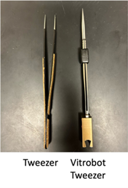
    Users may operate the Vitrobot based on level of training and experience (see Training and Access section below). Users must purchase their own set of tweezers specific to the Vitrobot from iCEM or from an approved vendor. 

  • Microscope Operation

    The Thermo Scientific Glacios can be used for both cryo-sample screening and cryo-data collection. 

    Operation 

    To maximize the amount of serviceable user time on the instruments, iCEM staff will perform operations for most users. This includes all grid clipping, loading, unloading, transfers, microscope alignments, low dose imaging, data collection, and troubleshooting. Following extensive training and approval by iCEM staff, some users may be allowed to perform some of these tasks independently (See Training and Access section). 

    Unforeseen Microscope Interruptions 

    The cryo-EM instrument is very complex with many potential points of failure. Unforeseen circumstances could potentially interrupt a user’s session. iCEM staff will do their best to minimize interruptions within their control. However, if a system failure does occur, the billed usage will be rounded to the nearest day.

    Users with an interrupted session will need to sign-up for a new session at the next available time. Please utilize iLab for this reservation or contact Mandy Bittner.

  • Microscope Performance and Quality Checks

    Beam alignment 

    The microscope will be aligned nand the image acquisition parameters will be adjusted by the iCEM staff before each session begins.

    Cryo-Cycle 

    A cryo-cycle is a necessary form of maintenance to expel water built up on cold surfaces. Cryo-cycling of the microscope will be performed once a week for 24 hours.  

    Data Processing 

    Users are to perform their own data processing and analysis. If desired, users are welcome to collaborate with the iCEM staff. The terms of such a collaboration would be the same as typical academic collaborations, which include contributing expertise and sharing authorship on publications and funding on grants. Due to the unpredictability of this process (in terms of the time needed and the potential end results), iCEM currently does not provide this as fee-for-service.

  • Training and Access

    Training will be provided with the goal of cultivating cryo-EM expertise in our community. Training for the use of electron microscope instruments will be carried out by the iCEM staff as authorized by the iCEM Director. Equipment training may be requested by contacting iCEM. Hands-on access to the instrumentation will be based on the level of training and experience of each user. The Director will decide when and whether a trainee has sufficient knowledge and proficiency to be designated normal, advanced, or expert user.

    A normal user must work on the microscope with the iCEM staff present. This user can only work during 9:00 am to 5:00 pm, Monday through Friday, with an approved iLab scheduling submission. Any overnight data collection must be ready by 5:00 pm. This user can perform sample preparation on the Vitrobot during regular business hours.

    An advanced user can work independently, meaning the user is certified to work weekends, holidays, and overnight. For certification, individual users must demonstrate proficiency in performing all required steps and tasks independently. Advanced users should be able to perform all necessary steps without having to refer to their notes and while operating all instrument components safely. This user can prepare samples on the Vitrobot, grid clipping, microscope operation, microscope alignment, setup EPU for data collection, and basic troubleshooting. An advanced user must still submit a scheduling request through iLab and this must be approved by iCEM staff.

    An expert user has the same access as an advanced user and may be qualified to load the cassettes into the microscope. Expert users are frequent and regular users of both the Glacios and Krios microscopes. An expert user must still submit a scheduling request through iLab and this must be approved by iCEM staff.

    Regardless of designation, all cassettes must be inspected by Dr. Yangshin Park before loading into the nano-capsule. This means that no sample can be loaded into the microscope without being first inspected by Dr. Park.