The In-Vivo Imaging Core provides imaging services for clinical trial, observational and preclinical studies. The core also provides radiochemistry support for molecular imaging. Currently, the Imaging Core is equipped with state-of-the-art imaging systems, including 3T MRI scanners, PET-CT scanners, CT scanners, Bruker 9.4T preclinical PET-MRI scanner, a Medical Cyclotron and Radiochemistry Facilities. Highly skilled faculty and staff are available to assist with research study designs.
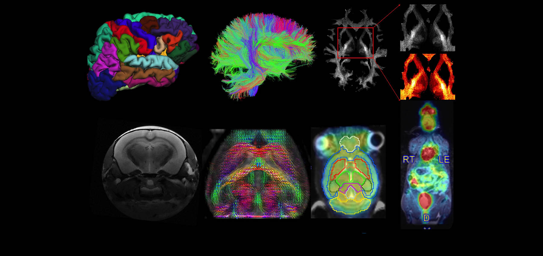
In-Vivo Imaging Core
Providing expert imaging services to further research and patient care throughout Indiana
147
active studies
$37 million
extramural funding support
7,000
imaging data


Services
-
MRINeuroimaging, cardiovascular imaging, musculoskeletal imaging, vasculature imaging and abdominal imaging. Charged by the hour with a 30-minute minimum. Contrast and other drugs charged separately.
-
PET-CTNeuroimaging, cardiovascular imaging and whole-body imaging. Charged by the hour with a 15-minute minimum. Radiopharmaceuticals, contrast. uptake time is charged separately.
-
Radiotracer Development and ProductionF-18( FDG, FET NaF, Fallypride , FSPG, MFBG, FET),N-13 Ammonia, AV-45,, C-11 (acetate, Raclopride, PIB, PBR28, Palmitate/GSK GSK‐1482160, UCB‐J) [O‐15]Water, Ga-PSMA-11. Please contact Scott Snyder, PhD for new Radiotracer development.
-
2023 Pricing
-
Research Scheduling
Scheduling of all research is to be made in advance with core personnel for the equipment being scheduled. Specific scheduling instructions will be provided after studies have been registered with the Office of Research Imaging (ORI). All users will be billed for the time scheduled OR for the time actually used, whichever is greater.
Cancellation and No Shows: Cancellation of scheduled scanner time must be two business days prior to the scheduled time or it will be considered a “no show” and billed at the full scan rate. If a radiopharmaceutical or other study drug is wasted due to a cancellation or no show the study will be charged for it. Late cancellations where the appointment slot gets filled will not be charged. -
Data Archival
Images are stored in Radiology’s long-term storage server. Study teams must specify if images are to be sent to a study specific server or outside destination.
Data should be removed from the scanner(s) as soon as possible after the examination. The imaging technologist has the right to remove all data older than one week. Data is the responsibility of the user (Principal Investigator). The In-Vivo Imaging Core may be contracted to archive data to specific locations for investigators; however, maintenance of the data is still the responsibility of the user.
-
Special Animal HandlingDepends on the availability of staff and upon request.
-
Imaging Technique DevelopmentIn the form of research collaboration.
-
Experimental DesignIn the form of research collaboration.
-
Data AnalysisIn the form of research collaboration.
Policies
-
Acknowledgment, Authorship, CollaborationPer CTSI policy, all imaging-related research work performed by the In-Vivo Imaging Core should be acknowledged in all ensuing publications. Fees paid for services provided by the core should not negate the qualification of co-authorship for the core scientists. These acknowledgements and achievements are important for the existence of the core facility. Given that many advanced imaging techniques and validated procedures require in-depth intellectual involvement by the core’s scientists (examples listed below), it is expected that this acknowledgement will be in the form of co-authorship according to the guidelines for authorship recommended by the International Committee of Medical Journal Editors. In-Vivo Imaging Core scientists performing one or more of the following tasks meet the criteria for authorship:
- Literature review and providing assistance in selecting/conceiving the experiments appropriate for the study goals
- Experimental design
- Imaging sequence development/optimization
- Data analysis and interpretation of data, or both
- Preparation of pertinent sections (e.g., methods) or figures for the manuscript and revisions to address reviewer concerns/questions
- Mere collection of image data using PI-provided protocols that require no further optimization and/or trouble-shooting.
- Mere collection of image study data without any data analyses or interpretation by the core scientists.
- Initial consultation.
- Assistance in image data transfer or storage.
- Clinical safety read for incidental findings
Please acknowledge the In-Vivo Imaging Core's help in a format similar to the one suggested below for publications and grant applications:
“… imaging was performed at the IIBIS In-Vivo Imaging Core, which was established and supported by the Department of Radiology and Imaging Sciences.”
Data obtained using facility equipment should include an acknowledgment of Indiana Institute for Biomedical Imaging Sciences.Text descriptions of the facility where equipment was used is available for researchers wishing to describe grant applications. The institute can provide letters of consultation or collaboration, as needed. It is requested that users of equipment supply the institute with an updated list every six months of publications and grant applications that referencing institute facilities.
-
ConfidentialityData generated by the institute is kept on a secure server with access to core personnel only.
-
Conflict ResolutionIf a resolution cannot be reached at the director level, the issue will be taken to an advisory committee for discussion and resolution.
-
Cost Recovery/Payment PolicyIf recovery of payment becomes an issue, business managers will contact the customer and/ or their department to collect charges owed. If there is a dispute in charges, the conflict resolution policy above will be followed.
-
Prioritization of Work
Studies or services will be on a first-come, first-serve basis. In the event that there is a back-log of studies or conflict in scheduling, services will be prioritized based on the following guidelines:
- Extramural Grant Funded Research
- Intramural and/or Industry Funded Research
- Standard of Care Diagnostic Scans Unrelated to Research
- Funded Pilot or Development Projects
- Unfunded Pilot or Development Projects
Facilities
Goodman Hall & Neuroscience building
R2 Building
Leadership

Yu-Chien Wu, MD, PhD
Associate Professor of Radiology & Imaging Sciences
Scientific Director

Scott E. Snyder, PhD
Associate Professor of Radiology & Imaging Sciences
Director, Molecular Imaging Ligand Development Program
